Animal Cell Diagram Under Light Microscope / B2 1 Cell Structure Igcse Aid / Draw a table of differences between the two this section on microscopy is meant as an introduction as learners will need to be able to use microscopes later.
byTrevor Bent-0
Animal Cell Diagram Under Light Microscope / B2 1 Cell Structure Igcse Aid / Draw a table of differences between the two this section on microscopy is meant as an introduction as learners will need to be able to use microscopes later.. Light microscopes using visible light and lenses to form a magnified image of the object under investigation e.g. It also has a very high resolving power. A cell is a very tiny structure which exists in living bodies. Image:plant cell seen under electron microscope. Light uses light waves as it's source of radiation and electron microscopes use electrons.
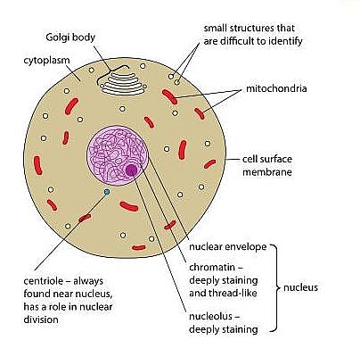
Image:animal cell seen under light microscope. A generalised animal cell as observed under an electron microscope. A cell is a very tiny structure which exists in living bodies. Nuclear pore in the last lesson you discussed cell membranes on the outside of. Line diagram of a general animal cell.
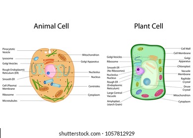
Section 5 Cells View As Single Page from www.open.edu Can you post a link for the cookie monster? Plant cells have cell walls, one large vacuole per cell, and chloroplasts, while animal cells will have a cell membrane only. Three main parts can be seen: Diagram 3.2 an animal cell. Animal cell labeled gif 572 417 pixels cell diagram science image information: Plant cell science diagram clipart set includes: Turn the coarse focus so that the stage is as close to the objective lens as possible. To use a light microscope to examine animal or plant cells.
The animal cell diagram on the free worksheet will teach students to identify the function of the major parts of the animal cell.
8 build a glossary of the cell parts. What was once unseeable can now be seen, touched, and eaten!cut. One hand goes under the base. Observing a wide range of biological processes and animal cell under light microscope is easier due to advances in microscopic techniques. Nuclear pore in the last lesson you discussed cell membranes on the outside of. To make observations and draw scale diagrams of cells. Describe and compare the structure of a plant cell with an animal cell, as seen under a light microscope, limited to cell wall, nucleus, cytoplasm, chloroplasts, vacuoles and location of the cell membrane. Chronic inflammation under the microscope learn share. See how a generalized structure of an animal cell and plant cell look with labeled diagrams. Light uses light waves as it's source of radiation and electron microscopes use electrons. Draw a diagram of one cheek cell and label its parts. Article was last reviewed on saturday, july 4, 2020. According to your observations in this lab and your book and notes, create a venn diagram to illustrate the structural similarities and differences between plant and animal cells.
We use microscope comprehensively in microbiology, mineralogy, cell biology, biotechnology, nano physics, microelectronics, pharmacology, and forensics. Anytime you are carrying your microscope, you should have two hands on it. Line diagram of a general animal cell. The golgi bodies are fairly easy to see under the microscope of many types of cells. Light microscopes using visible light and lenses to form a magnified image of the object under investigation e.g.
Plant Cell Nucleus High Res Stock Images Shutterstock from image.shutterstock.com .for viewing under the light microscope can label plant and animal cell structures and describe their functions to be able to work out the size of a cell 7 other animal cell features examiners tip: This is due to the lack of a cell wall. Preparing onion cell slides is a useful way to observe simple plant cells under the light microscope. Animal cell under a light microscope. Here's a photo of a plant cell under an electron microscope. We say cells are microscopic because they can only be seen under a microscope. Light microscope uses the properties of light to produce an enlarged image. Light uses light waves as it's source of radiation and electron microscopes use electrons.
Cells of plant or animal tissue.
This is due to the lack of a cell wall. Move the microscope to your workspace. The diagram is very clear, and labeled the diagram is very clear, and labeled; 720 x 1280 pixel type jpg download. Article was last reviewed on saturday, july 4, 2020. Study the two diagrams of plant and animal cells below. Describe and compare the structure of a plant cell with an animal cell, as seen under a light microscope, limited to cell wall, nucleus, cytoplasm, chloroplasts, vacuoles and location of the cell membrane. What was once unseeable can now be seen, touched, and eaten!cut. Here's a diagram of a plant cell: Light microscope uses the properties of light to produce an enlarged image. 417 x 572 pixel type jpg download. Plant cell science diagram clipart set includes: Light microscopes using visible light and lenses to form a magnified image of the object under investigation e.g.
Image:plant cell seen under electron microscope. It is much stronger than a light. Turn the coarse focus so that the stage is as close to the objective lens as possible. Structure of animal cell and plant cell under microscope diagrams image information: You should not look through the microscope to do this.
Animal Cell Structure And Organelles With Their Functions Jotscroll from www.jotscroll.com Line diagram of a general animal cell. Describe and compare the structure of a plant cell with an animal cell, as seen under a light microscope, limited to cell wall, nucleus, cytoplasm, chloroplasts, vacuoles and location of the cell membrane. Animal cell labeled gif 572 417 pixels cell diagram science image information: We use microscope comprehensively in microbiology, mineralogy, cell biology, biotechnology, nano physics, microelectronics, pharmacology, and forensics. Plant cells have cell walls, one large vacuole per cell, and chloroplasts, while animal cells will have a cell membrane only. Present to a significant degree in animal cells) to generate contrast. Diagram 3.2 an animal cell. What was once unseeable can now be seen, touched, and eaten!cut.
Image:animal cell seen under light microscope.
It is the simplest type of microscope. Cells consist of cytoplasm enclosed within a membrane, which contains many biomolecules such as proteins and nucleic acids.2 most plant and animal cells are only visible under a light microscope, with dimensions between 1 and 100 micrometres.3 electron microscopy gives a much higher. Resolving power is the ability to distinguish between separate things which are close to each other. See how a generalized structure of an animal cell and plant cell look with labeled diagrams. Study the two diagrams of plant and animal cells below. Chronic inflammation under the microscope learn share. Here's a photo of a plant cell under an electron microscope. But at the same time it is interpretive. Can you post a link for the cookie monster? Cell structure teaching resources the science teacher, organelles biology for majors i, 11 different types of cells in the human body, class test, chronic inflammation under the microscope learn share. Plant cells have cell walls, one large vacuole per cell, and chloroplasts, while animal cells will have a cell membrane only. Most of the cells are microscopic hence they can only be seen under a microscope in order to study their anatomy. Diagram 3.2 an animal cell.


Post a Comment